Laminitis Introduction

Information about laminitis in horses

Laminitis can be defined as inflammation of the sensitive laminae of the foot.

In order to better understand the mechanism of laminitis it is important to have a basic knowledge of the anatomy of the equine foot.

The pedal bone (third phalanx) is completely contained inside the hoof capsule. On the inside of the hoof there are insensitive (epidermal) laminae that interlink with sensitive (dermal) laminae present on the surface of the pedal bone. The weight of the horse is taken by the incredibly strong interlocking of these structures.

The deep digital flexor tendon has it attachment on the palmar surface (back) of the pedal bone. When the tendon pulls on the pedal bone, the whole of the hoof capsule is drawn into flexion, achieving movement of the distal limb.

During Laminitis there is a breakdown of the connection between epidermal and dermal laminae.

As a result, vertical displacement (sinking) of the pedal bone or rotation may occur due to the pulling force of the deep digital flexor tendon.

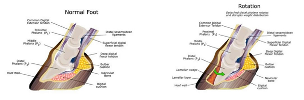
When rotation of the pedal bone occurs, abnormal pressure is applied to the sole, which becomes painful, especially at the toe.

Clinical Signs

Acute laminitis varies in severity and clinical signs include the following:


  • Increased digital pulses
  • Pain on hoof testers
  • “Pottery” gait
  • Sore in turning
  • Weight shifting at rest
  • Reluctance to move
  • Sweating and distress
  • Recumbency

A scale has been designed (Obel, 1948) to grade the severity of the laminitic episode:

  • Grade 1: Shifting weight at rest. Sound at walk. Short, stilted gait at trot
  • Grade 2: Stilted gait at the walk
  • Grade 3: Reluctant to move. Reluctant to have a foot picked up.
  • Grade 4: Refuses to move unless forced

Laminitis is still the subject of ongoing scientific research and many causes have been proposed:

  • Endotoxin-induced microthrombosis (but experimental administration of endotoxin failed to produce laminitis)
  • Alterations in Vascular dynamics: shunting of blood flow to foot, vasoconstriction or vasodilation
  • Enzyme (Metalloproteinase) destruction of attachment between hoof wall and pedal bone due to inappropriate release of activated metalloproteinase

While a definitive cause has not been yet found, several trigger factors have been identified:

The diagnosis of laminitis is based on:

  • Clinical examination (lameness, presence of digital pulse, painful response on hoof testers, dip in coronary band, heart rate)
  • Radiographic examination to evaluate degree of rotation/drop of pedal bone (dislocation) and establish prognosis and treatment options

Therapy

The therapy in the acute phase (immediately) is aimed to:

  • Stabilise pedal bone (prevent further rotation/drop) : Box rest on deep bedding (preferably shavings),frog support, toe trimming (if comfortable enough), removal of shoes (if comfortable enough)
  • Decrease pain and inflammation of the laminae (administration of non-steroidal anti-inflammatory drugs like Flunixin Meglumine, Phenylbutazone, Suxibuzone)
  • Keep horse calm and increase peripheral blood flow (Acepromazine)
  • Treatment of concurrent pathologies (e.g. Metritis, retained placenta, endotoxemia)
  • Tubing with laxatives in cases of feed shed burglary
  • Percutol patches in cases of feed shed burglary

The therapy in the subacute phase (after 1-2 weeks) is aimed to maximise comfort and to support the pedal bone. X-rays are very useful to assess the position of the pedal bone inside the hoof capsule and to assist in therapeutic trimming and shoeing (dorsal wall resection, glue-on shoes). The management of the chronic laminitic should be aimed to:

  • Keep weight under control (Muzzle if grass lush/ restrict grazing, minimal amounts of hard feed)
  • Low sugar, low starch feed
  • Keep feet balanced with regular trimming
  • Treat any concurrent predisposing pathologies (PPIDEMS)
Return to Equine Information